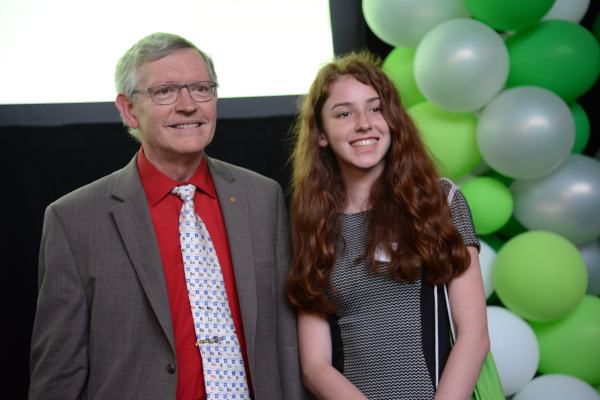
The Keynote Address for the 2017 California State Science Fair was presented by Dr. W.E. Moerner, the Harry S. Mosher Professor of Chemistry and Professor by courtesy of Applied Physics at Stanford University, and a recipient of the Nobel Prize in Chemistry, 2014. The following presentation was provided to CSSF by Dr. Moerner in lieu of a transcript from the Keynote Address. This presentation is not identical to the Keynote Address, but does cover the same essential points, tells the story behind his Nobel Prize, and includes a bit of early life history. We are grateful to Dr. Moerner for allowing us reproduce to this presentation.
In 2014, along with Eric Betzig and Stefan Hell, I received the Nobel Prize in Chemistry for something called super-resolution fluorescence microscopy. It sounds like a bit of a mouthful, but let me explain how I was involved, and why it matters to you more than you might think — and what our path to the breakthrough reveals about the nature of scientific research today.
When I was growing up, I simply loved learning! There was something special about math and science in the way they give us a way to understand how the world works. I had a great time with experiments, such simple chemical reactions from a chemistry set, performed in a metal house in my back yard, and disassembling every resistor and capacitor I could find in those old, huge tube-style TV sets, even though I dropped a little bit of hot solder on my leg now and then! Of course, I made mistakes now and then, such as working on the washing machine in bare feet (ouch!), but the key is to always learn from your mistakes. I became an amateur (“ham”) radio operator in high school along with my father, and the thrill of radio (hearing the unseen) and the beauty of electromagnetic waves played a large part in the Nobel Prize work! It was after I finished college and graduate school, that while working at IBM Research Labs, my postdoc Lothar Kador and I first used light and electromagnetic waves to optically detect a single molecule, almost 30 years ago (1989). This was a wonderful achievement, because seeing one molecule meant that we no longer had to compute averages all the time — we can watch single molecules one at a time to see if they are “marching to different drummers” or not! This was a key step toward the Nobel Prize, but there is more to the story.
Now to set the stage: Before microscopes were first invented a few hundred years ago, the smallest thing we could see was about the width of a human hair. The invention of the microscope literally opened up whole new worlds for scientific research, such as seeing tiny organisms that can lead to illness like bacteria. But there were still serious limits on the smallest of scales. Almost everything in our world is made of molecules. But molecules are extremely tiny — just a few nanometers in size. Remembering that one human hair is about 100 micrometers in diameter, a nanometer is 100,000 times smaller! Many proteins and enzymes in cells that make biology work are around 10 nanometers in size. These objects are so small that when viewed through a microscope, the resulting image looks blurry and out of focus, no matter how expensive the microscope, due to the “diffraction limit” of light.
What exactly is the diffraction limit? When scientists are trying to observe something, we’ll often take fluorescent (light-emitting) molecules and attach them only to the structure of interest, because that way, the light we see only comes from the object we want to observe. But here’s the problem: the wavelength of visible light is roughly 500 nanometers, which in the case of molecules, is much, much bigger than the structures we want to see. For microscopes, this means that an image of a single 1-nanometer-sized emitting molecule looks far, far larger than 1 nanometer. In fact, a single molecule emitting light looks like something that is 250 nanometers in size, let’s call it a fuzzball!
So when scientists tried to view molecules or structures in cells that are only a few nanometers in size using conventional optical microscopes, they always got very fuzzy pictures because the fuzzballs from the molecules would all overlap. That was a serious, fundamental problem. The main goal of science is to understand how things work. For example, in cell biology, we want to understand in every exquisite detail exactly how the tiny “nanomachines” inside cells work. If we learn more about how normal cells work, we can apply the same methods to diseased cells, and this can ultimately help us correct things when they go wrong. But how can we do that if we can’t even see the things we want to study? This problem plagued optical microscopy since its beginnings.
Let’s go back to 1989, when single molecules were first observed with light as I mentioned above. Our first experiment was fairly esoteric in that it required extremely low temperatures near absolute zero, and we were basically exploring the ultimate single-molecule limit because no one had done this before. Many scientists all over the world started similar experiments and there were great steps forward by people like Michel Orrit in France. But we realized our work was going to be important when we observed that single, individual molecules were doing some amazing things. Individual molecules would jump from one color or wavelength to another! Or they would turn on and off, and blink in interesting ways!
It is because molecules do this — have an individual on/off behavior — that we were able to start chipping away at a long-held belief in the scientific community: that we would never be able to observe things smaller than around half the wavelength of light. Microscopes could now become nanoscopes.
So how did the initial observation of single molecules, and the discovery of single-molecule blinking, pave the way for nanoscopes that could bring even tiny molecules into sharp focus?
Because, as scientists like Eric Betzig and others realized in 2005, we could remove the fuzziness by choosing emitting molecules that had two states: an “on” state where light is emitted, and an “off” state, where light is not emitted, and most importantly, image them at different times. To see this, imagine we place our molecules along the structure we want to image, and we only turn a few “on” at any time. We then film them. In the different frames of the movie, the sample looks like stars in the sky or fireflies at night, and because the different spots are separated, we can determine the position of each molecule very precisely. The key point is that the molecules randomly turn on and off. Just by watching them, then over time, we accumulate a long list of the positions of the molecules, and then we use computer graphics to show them all at once, like pointillism in art. The structure we wanted to see is now much sharper than was possible before! A fundamental limit since the start of microscopy had been surpassed.
As an approximate analogy, suppose you wanted to see the branches of a tree, but it is dark. What you could do is place fireflies along the branches of the tree, then just let them blink randomly on and off. Every time you see one turn on, you carefully measure its exact position. Even if you used an out-of-focus camera, you can still find the center of each spot from each firefly. Then you take all the positions you found, show them all at once, and the branches appear!

The implications of this breakthrough are enormous: now, the focus of microscopes can be improved by five times, ten times, even 100 times so that extremely tiny structures can be observed. Scientists have already used the technology to see cell structures that were never visible before, from microtubules to protein aggregates that occur in Parkinson’s, Alzheimer’s and Huntington’s diseases. Much remains to be done to refine this idea and apply it in fields ranging from biology to materials science.
I had no idea, back in 1989, that the work I was doing would eventually lead to such a breakthrough that would open up whole new research areas. But without the desire to achieve something beyond what was possible before — and the funding that supported this type of “what if” basic research — we would not be where we are today. Exploring the unexplored can lead to life-changing applications that cannot be predicted at the start, and our world depends upon this kind of research to build our future.
After the Opening Ceremony concluded Dr. Moerner graciously met with many of the students who were present at the Keynote Address. A few of them are pictured below.

